Showing posts with label Skull seen from the front.. Show all posts

Wednesday, July 11, 2012

The Hyoid Bone


Fig. 51.
Hyoid bone, seen from the front.

Fig. 52.
Hyoid bone seen from the lateral side.

Fig. 53.
Median section through the hyoid bone (body).

Fig. 54.
Attachments on the hyoid bone seen from the anterosuperior aspect.

Fig. 55.
Schematic parasagittal section through the body of the hyoid bone, to show the arrangement  of structures attached.

Fig. 56.
Schematic vertical section through a greater cornu of the hyoid bone to show the arrangement of structures attached.

The Mandible


Fig. 43.
The mandible seen from above.

Fig. 44.
Right half of mandible seen from the lateral side.

Fig. 45.
Right half of the mandible seen from the medial side.

Fig. 46.
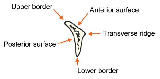
Median part  of the mandible, anterior aspect.

Fig. 47.
Median part of the mandible, posterior aspect.

Fig. 48.
Attachments on the mandible as seen from the lateral side.

Fig. 49.
Attachments on the mandible as seen from the medial side.

Fig. 50.
Region of the symphysis menti, showing attachments, seen from behind.

Tuesday, July 10, 2012

The Nasal Cavity


Fig. 37A.
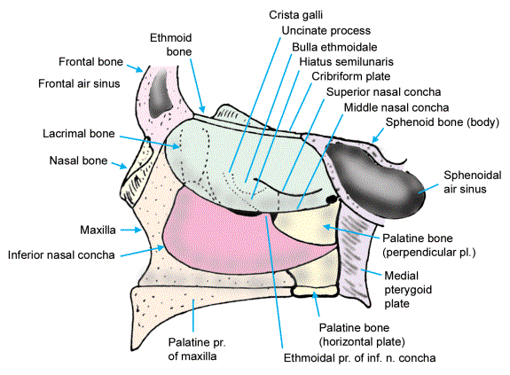
Bones in the lateral wall of the nose. The base of the lateral wall formed by the maxilla.

Fig. 37B.
Palatine bone overlaps the  posterior part of the maxilla.

Fig. 37C.
The lacrimal bone and the inferior nasal concha are placed over the maxilla and the palatine bone.

Fig. 38.
Lateral wall of the nasal cavity is completed by the ethmoid bone. The parts shown in dotted line can be seen only when the middle concha is lifted off. Compare with Fig. 37C.

Fig. 39.
Main bones forming the nasal septum.

Fig. 40.
Lateral wall of the nasal cavity seen after removal of the medial plate of the ethmoid bone to expose the ethmoidal air sinuses.


Fig. 41.
Schematic diagram to show the positions of the openings of paranasal sinuses into the nasal cavity.

Fig. 42.
Scheme to show the parts of the   ethmoid bone.


Foramina Of The Skull


Fig. 31.
Structures passing through the optic canal, the superior orbital fissure, and the inferior orbital fissure.

Fig. 32.
Structures passing through the foramen ovale, and through smaller foramina near it.
The lesser petrosal nerve sometimes passes through the canalis innominatus
.

Fig. 33.
Scheme to show structures passing through the carotid canal and the foramen lacerum.

Fig. 34.
Scheme to show structures passing through the jugular foramen.

Fig. 35.
Scheme to show the position of the opening of the inferior tympanic canaliculus,
and of the mastoid canaliculus.

Fig. 36.
Scheme to show the arrangement of structures passing  through the foramen magnum.

Monday, July 9, 2012

Cranial Fossae Of The Skull


Fig. 28.
Parts of the anterior and middle cranial fossae seen from above. Compare with Fig. 7.

Fig. 29.
Schematic diagram to show some features in the anterior wall of the middle cranial fossa.

Fig. 30.
Features to be seen in the floor of the middle and posterior cranial fossae

Wednesday, July 4, 2012

Features On The Skull As Seen From The Lateral Side


Fig. 14.
Sutures seen on the lateral aspect of the skull.

Fig. 15.
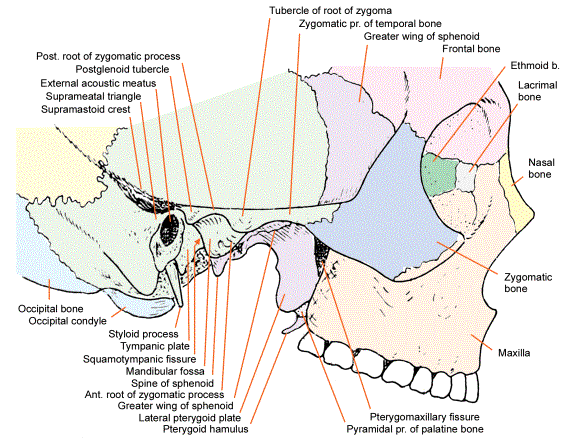
Some features seen on the lateral aspect of the skull. Also see Figs. 1, 5, 14, 16 & 17.

Fig. 16.
Additional features seen on the lateral aspect of the skull. Also see Figs. 1, 5, 14, 15 and 17.

Fig. 17.
Some details seen on the lateral aspect of the skull after removing the zygomatic arch.

Fig. 18.
Anterior wall of temporal and infratemporal fossae seen from behind. The zygomatic process of the temporal bone has  been removed and a coronal section cut through the middle cranial fossa

Fig. 19.
Scheme to show the walls of the pterygopalatine fossa

Fig.20.
Attachments on the skull seen from the lateral side.

Monday, July 2, 2012

Features On The Skull Seen From Above and Behind


Fig. 12.
Features seen on the skull when viewed from above.
Compare with Fig. 2.

Fig. 13.
Features seen on the skull when viewed from behind.
Compare with
Fig. 3.

Features On The Front Of The Skull


Fig. 8.
Some details of features of the skull as seen from the front. Compare with Figs. 4 and 11.

Fig. 9.
Schematic coronal section through the orbit and nasal cavity to show the bones related to them.

Fig. 10. Attachments on the anterior aspect of the skull.

Fig. 11.
The orbit and surrounding structures. The figure is schematic to the extent that all
features shown cannot be seen from one fixed angle of viewing.