Showing posts with label Foramina of skull. Show all posts

Monday, July 23, 2012

Inflammation and Repair 1 Outline (part-2) In Pathologic Basis Of Disease


6 - Lung, fibrinous and hemorrhagic inflammation, pneumonia - High power (Glass slide 41)
  • Congested capillaries in alveolar walls
  • Erythrocytes
At this higher power, the nature of the inflammatory exudate is clearer. It consists of erythrocytes that have leaked out of the very permeable and congested capillaries, thin strands of fibrin derived from fibrinogen in the exuded plasma, and leukocytes that have exited the vessels via diapedesis. 

7 - Hand, purulent inflammation, staphylococcal abscess - Clinical presentation
  • Abscess
Inflammation involving a dense neutrophilic infliltrate is called purulent (referring to pus) or suppurative. A localized collection of neutrophils, necrotic cells, and edema fluid (pus) in tissue is an abscess. Collections of neutrophils tend to have a greenish-yellow or whitish-yellow color, as seen here.


8 - Trachea, bronchi and lungs, purulent inflammation, bronchopneumonia and abscess - Gross, cut surface
  • Abscess
  • Purulent inflammation 

Note the pale yellow color of the area of purulent inflammation. In one focus, this inflammation has formed an abscess.


9 - Lung, purulent inflammation, abscess - Very low power (Glass slide 46)

  • Abscesses
In this section of the lung, the large, irregularly shaped defects lined by shaggy, basophilic material are abscesses. They appear empty, because the liquid purulent material washed away when the tissue was sectioned to prepare the slide. The remaining parenchyma appears consolidated.

10 - Lung, purulent inflammation, abscess - High power (Glass slide 46)
This high-power view of the wall of the abscess demonstrates the dense neutrophilic infiltrate.

Wednesday, July 11, 2012

The Hyoid Bone


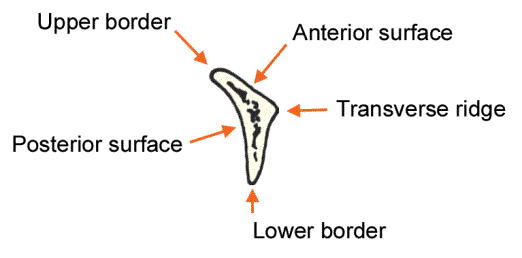
Fig. 51.
Hyoid bone, seen from the front.

Fig. 52.
Hyoid bone seen from the lateral side.

Fig. 53.
Median section through the hyoid bone (body).

Fig. 54.
Attachments on the hyoid bone seen from the anterosuperior aspect.

Fig. 55.
Schematic parasagittal section through the body of the hyoid bone, to show the arrangement  of structures attached.

Fig. 56.
Schematic vertical section through a greater cornu of the hyoid bone to show the arrangement of structures attached.

The Mandible


Fig. 43.
The mandible seen from above.

Fig. 44.
Right half of mandible seen from the lateral side.

Fig. 45.
Right half of the mandible seen from the medial side.

Fig. 46.
Median part  of the mandible, anterior aspect.

Fig. 47.
Median part of the mandible, posterior aspect.

Fig. 48.
Attachments on the mandible as seen from the lateral side.

Fig. 49.
Attachments on the mandible as seen from the medial side.

Fig. 50.
Region of the symphysis menti, showing attachments, seen from behind.

Tuesday, July 10, 2012

The Nasal Cavity


Fig. 37A.
Bones in the lateral wall of the nose. The base of the lateral wall formed by the maxilla.

Fig. 37B.
Palatine bone overlaps the  posterior part of the maxilla.

Fig. 37C.
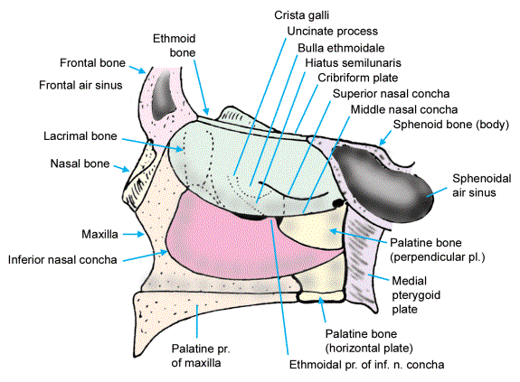
The lacrimal bone and the inferior nasal concha are placed over the maxilla and the palatine bone.

Fig. 38.
Lateral wall of the nasal cavity is completed by the ethmoid bone. The parts shown in dotted line can be seen only when the middle concha is lifted off. Compare with Fig. 37C.

Fig. 39.
Main bones forming the nasal septum.

Fig. 40.
Lateral wall of the nasal cavity seen after removal of the medial plate of the ethmoid bone to expose the ethmoidal air sinuses.


Fig. 41.
Schematic diagram to show the positions of the openings of paranasal sinuses into the nasal cavity.

Fig. 42.
Scheme to show the parts of the   ethmoid bone.


Foramina Of The Skull


Fig. 31.
Structures passing through the optic canal, the superior orbital fissure, and the inferior orbital fissure.

Fig. 32.
Structures passing through the foramen ovale, and through smaller foramina near it.
The lesser petrosal nerve sometimes passes through the canalis innominatus
.

Fig. 33.
Scheme to show structures passing through the carotid canal and the foramen lacerum.

Fig. 34.
Scheme to show structures passing through the jugular foramen.

Fig. 35.
Scheme to show the position of the opening of the inferior tympanic canaliculus,
and of the mastoid canaliculus.

Fig. 36.
Scheme to show the arrangement of structures passing  through the foramen magnum.